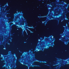
Macrophages provide a marker for mild TBI diagnosis
Interview with
TBI diagnostics are still pretty rudimentary. MRI scans are useful for spotting structural changes to the brain - lesions, for example, which might be an indication of severe TBI - but fail to detect the more subtle changes in brain functionality associated with mild traumatic brain injury.
A team of researchers at Harvard University have been working on a solution. Using part of the body’s immune response to head trauma, they’ve found a biomarker which they can take advantage of to ensure fewer mild TBIs go undiagnosed. They’re called macrophages. Professor Samir Mitragotri explains…
Samir - So macrophages are the body's immune cells. They are among the most common immune cells, and what is unique about them is that whenever there is damage or inflammation in tissue, they infiltrate into the tissue and they become a part of the tissue. We figured that even though the trauma may be subtle, that the structure will not be visibly changed, the macrophages may know that the trauma has happened and may infiltrate into the brain and maybe we can use that signal to detect the extent of trauma.
James - Could they be getting to the brain for any other reason other than a TBI?
Samir - That's quite possible. Macrophages chase inflammation. If there are other reasons to go into the brain, they very well might. But in the case of TBI, if we suspect that the brain has suffered a trauma, which is typically what happens when you bring in a subject: because a fall has happened, a trauma has happened, in that case, if you see the inflammation, if you see the macrophages going into the brain, that is a good way to see whether the trauma has happened.
James - I see. Where did the idea come from to use macrophages in TBI diagnostics? What was the leap there?
Samir - So the leap to use macrophages really comes from the physiology. The challenge has been how do you see them? They're not visible under MRI. We wanted to attach a contrast agent to them so that we can track these macrophages. That turned out to be quite a challenge because macrophages are the bodies professional eaters, they will basically eat whatever they bind to. In fact, that's their job. So how do you attach something to the macrophage without having it eaten by the macrophage? We had made a discovery a while ago that macrophages cannot internalise disc shaped particles when they bind to them. So we figured out a way to make a disc shaped backpack and we put gadolinium in it.
James - This is gadolinium, a commonly used contrast agent, a substance which will light up on an MRI like a Christmas tree, which you were able to put in the disc shaped hydrogel backpack you developed worn by macrophages as they travel to the brain to fight inflammation from a mild TBI.
Samir - Indeed. And that's what we call a GLAM, which is basically a micron sized hydrogel disc that is loaded with a high concentration of gadolinium, and that becomes our tracker of the macrophage, wherever it is in the body. Gadolinium is a small molecule entity, so when you inject in the blood, it is cleared pretty quickly, within minutes from the body. And in fact, it is cleared by kidney filtration. That's been one of the challenges. Those patients who may be suffering from kidney malfunctioning, they cannot really use the current version of gadolinium very effectively. So when it came to putting gadolinium in our backpacks, two constraints had to be met: one is that gadolinium needs to make contact with water because that's the mechanism by which it provides a contrast. So to allow the water molecules to come in close contact with gadolinium, we went with the hydrogel design for the backpack. It's very porous so water molecules can come in. And the second design factor was that because hydrogels are porous, gadolinium can potentially leak out. To stop that from happening, we covalently conjugated gadolinium to the backpack. And to make that happen, we had to make a special version of the gadolinium molecule which can be incorporated into the backpack. By doing these two modifications, we were able to make backpacks which can provide a strong enough contrast in MRI.
James - And did they work? Have you tested the effectiveness of your GLAM hitchhikers and what were the results?
Samir - They did work. So we tested the GLAMs in a pig model. That work was done in collaboration with a researcher at Boston Children's Hospital. The way this was tested is that mild TBI was induced in pigs, and when we injected these animals with GLAMs, we saw that we are able to see the occurrence of mild TBI under the conditions where the conventional gadolinium contrast could not provide an indication.
James - An incredible bit of science. Is there the potential for this sort of work to be extrapolated into other diagnostic tools for us to use the immune system to help to diagnose other diseases?
Samir - We believe so. At the heart of it, we are essentially tracking the motion of macrophages and macrophages are very sensitive indicators of inflammation and injury. So by being able to see where they are and how they're moving about, I think one can make a very sensitive and very differentiated diagnosis of many other conditions. What we have done in this study is used it to detect mild TBI, but we do believe that there is potential to extrapolate these two other indications and the research will have to be done to get a better assessment as to what those indications would be and how would this method be superior compared to the current standards for those diseases.


Comments
Add a comment